L’imagerie médicale au service de l’impression 3D / Medical imaging is employed by 3D printing
Théophraste Lescot, groupe de recherche de Marc-André Fortin, Canon Rebel T5i – MeshLab
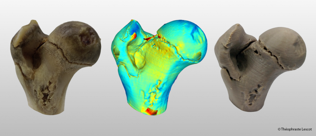
L’image de gauche est la partie supérieure d’un fémur de porc. Après avoir été imagé à l’aide de la tomodensitométrie par rayon X (TDM-X), des traitements numériques génèrent le modèle 3D de l’os de porc. L’image de droite est une impression 3D en polyétheréthercétone (PEEK) du modèle 3D de l’os de porc. Après avoir été imagé par TDM-X aussi, un modèle 3D de l’impression en PEEK est obtenu. L’image centrale est une représentation 3D de la conformité géométrique entre le modèle imprimé en 3D et l’os. Les couleurs chaudes représentent un surplus de matière, tandis que les couleurs froides correspondent à un manque de matière.
—————————————-
The image on the left is the upper part of a pig femur. After being imaged using X-ray computed tomography (X-CT), digital processing generates the 3D model of the pig bone. The image on the right is a polyetheretherketone (PEEK) 3D printed part of the 3D pig bone model. After being imaged by X-CT as well, a 3D model of the PEEK printed part is obtained. The central image is a 3D representation of the geometrical conformity between the 3D printed model and the bone. Warm colors represent an excess of material, while cold colors correspond to a lack of material.